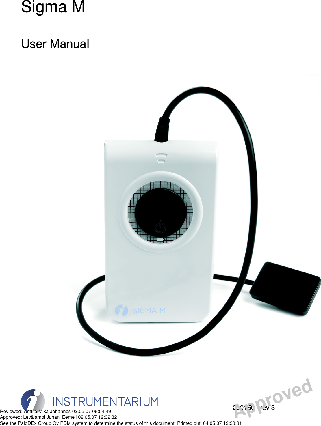
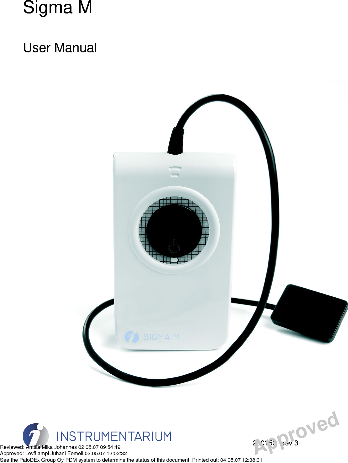
Instrumentarium Dental PaloDEx Group SM Digital Intraoral Sensor User Manual
Instrumentarium Dental, PaloDEx Group Oy Digital Intraoral Sensor
UserManual.wiki
>
Instrumentarium Dental PaloDEx Group
>
SM User Manual
User manual
Navigation menu
Upload a User Manual
Namespaces
Wiki Guide
HTML
PDF
Info
Views
User Manual
Discussion / Help
Navigation


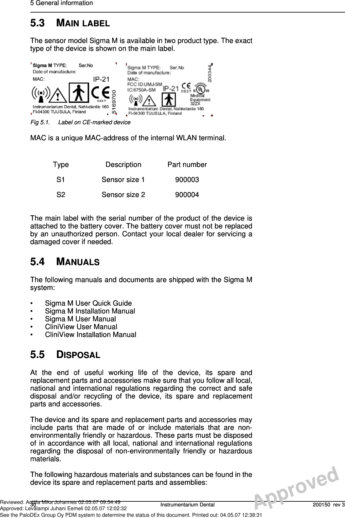
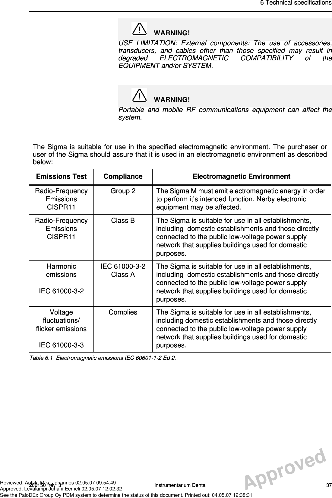
![6 Technical specifications38 Instrumentarium Dental 200150 rev 3Recommended Separation Distances for Portable and Mobile RF Communications Equipment IEC 60601-1-2Frequency of TransmitterEquationRated Maximum Output Power of Transmitter (watts)150KHz to 80 MHzSeparation Distance (meters)80 MHz to 800 MHzSeparation Distance(meters)800 MHz to 2,5 GHzSeparation Distance(meters)0.01 0.12 0.12 0.230.1 0.37 0.37 0.741 1.17 1.17 2.3410 3.69 3.69 7.38100 11.67 11.67 23.34Table 6.2 Recommended Separation Distances for Portable and Mobile RF Communications Equipment IEC 60601-1-2 Ed 2.PV = d]5,3[1PE = d]5,3[1PE = d]7[16 Technical specifications38 Instrumentarium Dental 200150 rev 3Recommended Separation Distances for Portable and Mobile RF Communications Equipment IEC 60601-1-2Frequency of TransmitterEquationRated Maximum Output Power of Transmitter (watts)150KHz to 80 MHzSeparation Distance (meters)80 MHz to 800 MHzSeparation Distance(meters)800 MHz to 2,5 GHzSeparation Distance(meters)0.01 0.12 0.12 0.230.1 0.37 0.37 0.741 1.17 1.17 2.3410 3.69 3.69 7.38100 11.67 11.67 23.34Table 6.2 Recommended Separation Distances for Portable and Mobile RF Communications Equipment IEC 60601-1-2 Ed 2.PV = d]5,3[1PE = d]5,3[1PE = d]7[1ApprovedReviewed: Anttila Mika Johannes 02.05.07 09:54:49Approved: Levälampi Juhani Eemeli 02.05.07 12:02:32See the PaloDEx Group Oy PDM system to determine the status of this document. Printed out: 04.05.07 12:38:31](https://usermanual.wiki/Instrumentarium-Dental-PaloDEx-Group/SM/User-Guide-825850-Page-44.png)
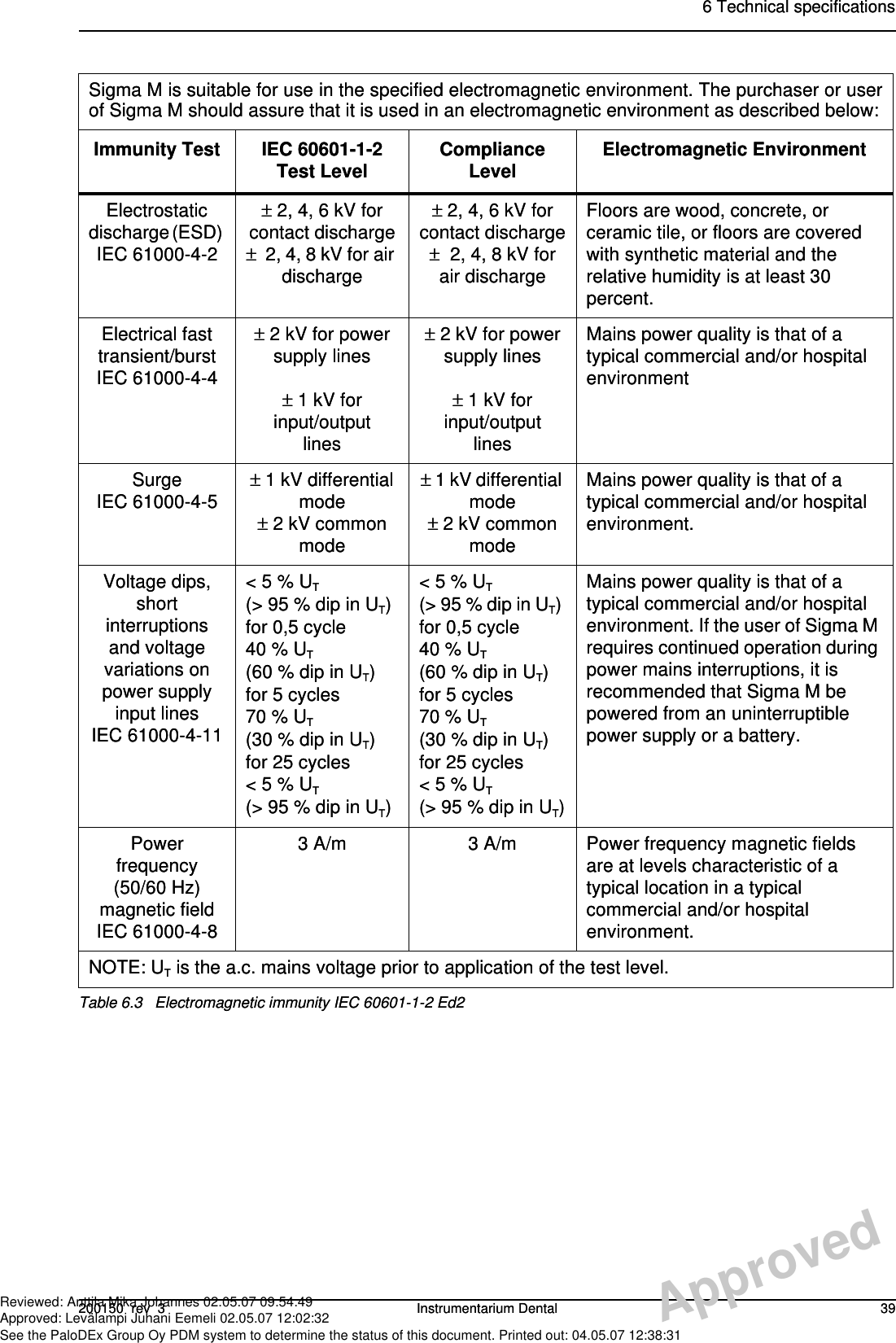
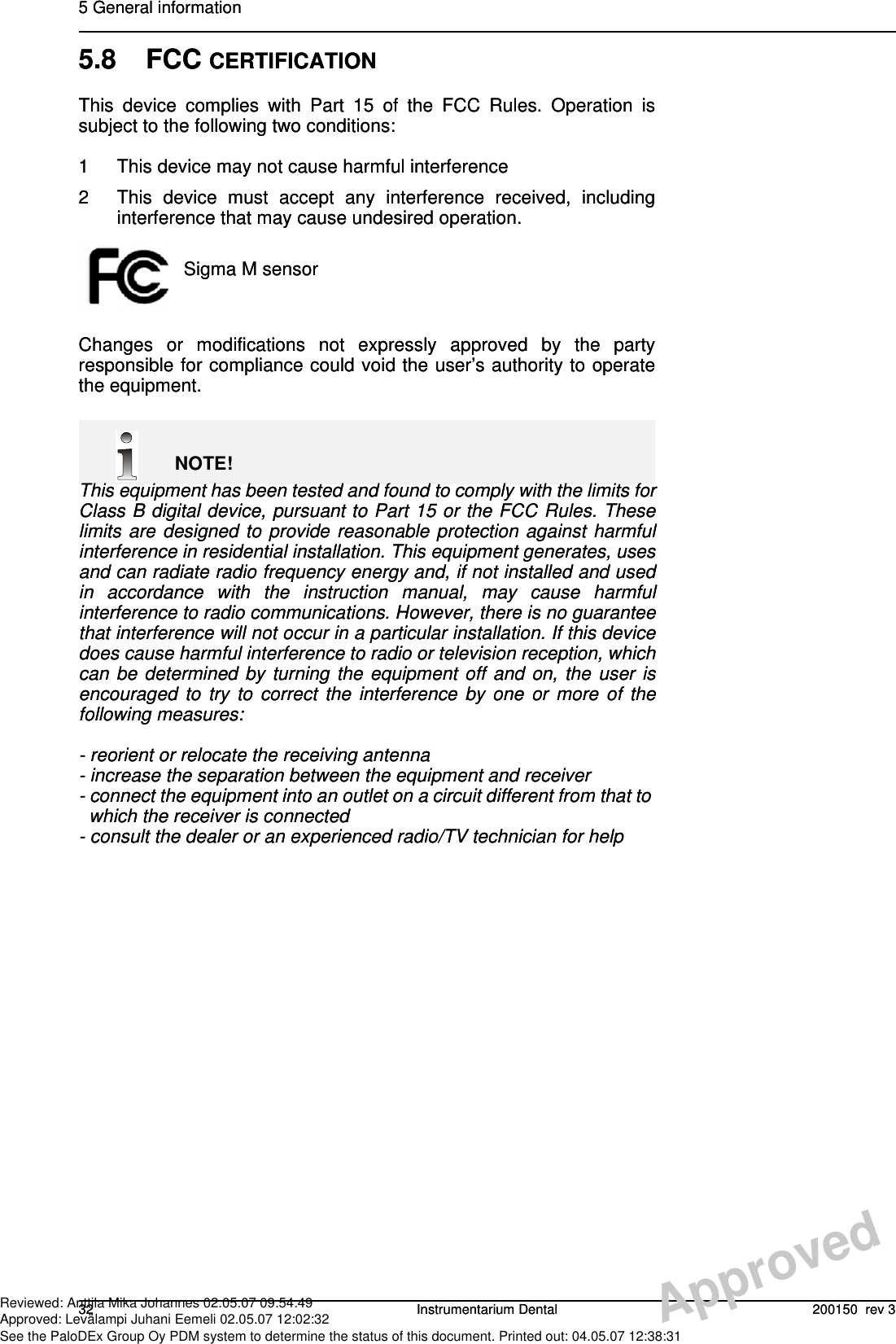
![6 Technical specifications40 Instrumentarium Dental 200150 rev 3Sigma M is suitable for use in the specified electromagnetic environment. The purchaser or userof Sigma M should assure that it is used in an electromagnetic environment as described below:ImmunityTest IEC60601-1-2Test LevelComplianceLevel ElectromagneticEnvironmentConducted RF IEC 61000-4-6Radiated RFIEC 61000-4-33 V150 kHz to80 MHz3 V/m80 MHz to2,5 GHz[ V1 ] 3 V[ E1 ] 3 V/mPortable and mobile RF communications equipment are used no closer to any part of Sigma M, including cables, than the recommended separation distance calculated from the equation appropriate for the frequency of the transmitter.Recommended Separation Distance: 80 MHz to 800 MHz 800 MHz to 2,5 GHzWhere P is the maximum output power rating of the transmitter in watts (W) according to the transmitter manufacturer and d is the recommended separation distance in meters (m).Field strengths from fixed RF transmitters, as determined by an electromagnetic site survey,* are less than the compliance level in each frequency range.** Interference may occur in the vicinity of equipment marked with the following symbol: *Field strengths from fixed transmitters, such as base stations for cellular telephones and landmobile radios, amateur radio, AM and FM radio broadcast, and TV broadcast cannot beestimated accurately. To assess the electromagnetic environment due to fixed RF transmitters,an electromagnetic site survey should be performed. If the measured field strength exceeds theRF compliance level above, observe Sigma M to verify normal operation in each use location. Ifabnormal performance is observed, additional measures may be necessary, such as re-orientingor relocating Sigma M.**Over the frequency range 150 kHz to 80 MHz, field strengths are less than [V1] V/m.The Recommended Separation Distances are listed on page 21.Note: These guidelines may not apply in all situations. Electromagnetic propagation is affectedby absorption and reflection from structures, objects, and people.Table 6.4 RF immunity of non-life-support equipment or system IEC 60601-1-2PV = d]5,3[1PE = d]5,3[1PE = d]7[16 Technical specifications40 Instrumentarium Dental 200150 rev 3Sigma M is suitable for use in the specified electromagnetic environment. The purchaser or userof Sigma M should assure that it is used in an electromagnetic environment as described below:ImmunityTest IEC60601-1-2Test LevelComplianceLevel ElectromagneticEnvironmentConducted RF IEC 61000-4-6Radiated RFIEC 61000-4-33 V150 kHz to80 MHz3 V/m80 MHz to2,5 GHz[ V1 ] 3 V[ E1 ] 3 V/mPortable and mobile RF communications equipment are used no closer to any part of Sigma M, including cables, than the recommended separation distance calculated from the equation appropriate for the frequency of the transmitter.Recommended Separation Distance: 80 MHz to 800 MHz 800 MHz to 2,5 GHzWhere P is the maximum output power rating of the transmitter in watts (W) according to the transmitter manufacturer and d is the recommended separation distance in meters (m).Field strengths from fixed RF transmitters, as determined by an electromagnetic site survey,* are less than the compliance level in each frequency range.** Interference may occur in the vicinity of equipment marked with the following symbol: *Field strengths from fixed transmitters, such as base stations for cellular telephones and landmobile radios, amateur radio, AM and FM radio broadcast, and TV broadcast cannot beestimated accurately. To assess the electromagnetic environment due to fixed RF transmitters,an electromagnetic site survey should be performed. If the measured field strength exceeds theRF compliance level above, observe Sigma M to verify normal operation in each use location. Ifabnormal performance is observed, additional measures may be necessary, such as re-orientingor relocating Sigma M.**Over the frequency range 150 kHz to 80 MHz, field strengths are less than [V1] V/m.The Recommended Separation Distances are listed on page 21.Note: These guidelines may not apply in all situations. Electromagnetic propagation is affectedby absorption and reflection from structures, objects, and people.Table 6.4 RF immunity of non-life-support equipment or system IEC 60601-1-2PV = d]5,3[1PE = d]5,3[1PE = d]7[1ApprovedReviewed: Anttila Mika Johannes 02.05.07 09:54:49Approved: Levälampi Juhani Eemeli 02.05.07 12:02:32See the PaloDEx Group Oy PDM system to determine the status of this document. Printed out: 04.05.07 12:38:31](https://usermanual.wiki/Instrumentarium-Dental-PaloDEx-Group/SM/User-Guide-825850-Page-46.png)